


The main LPAW clinic is in Bow, E3, London, right next to The Bow Quarter. This bright and spacious clinic offers 4 treatment rooms, 2 changing rooms with showers, a large rehab gym, & onsite hydrotherapy in our 17 foot pool.
The LPAW satellite clinic is based in Stratford East Village where we run a thriving sports rehab offering.
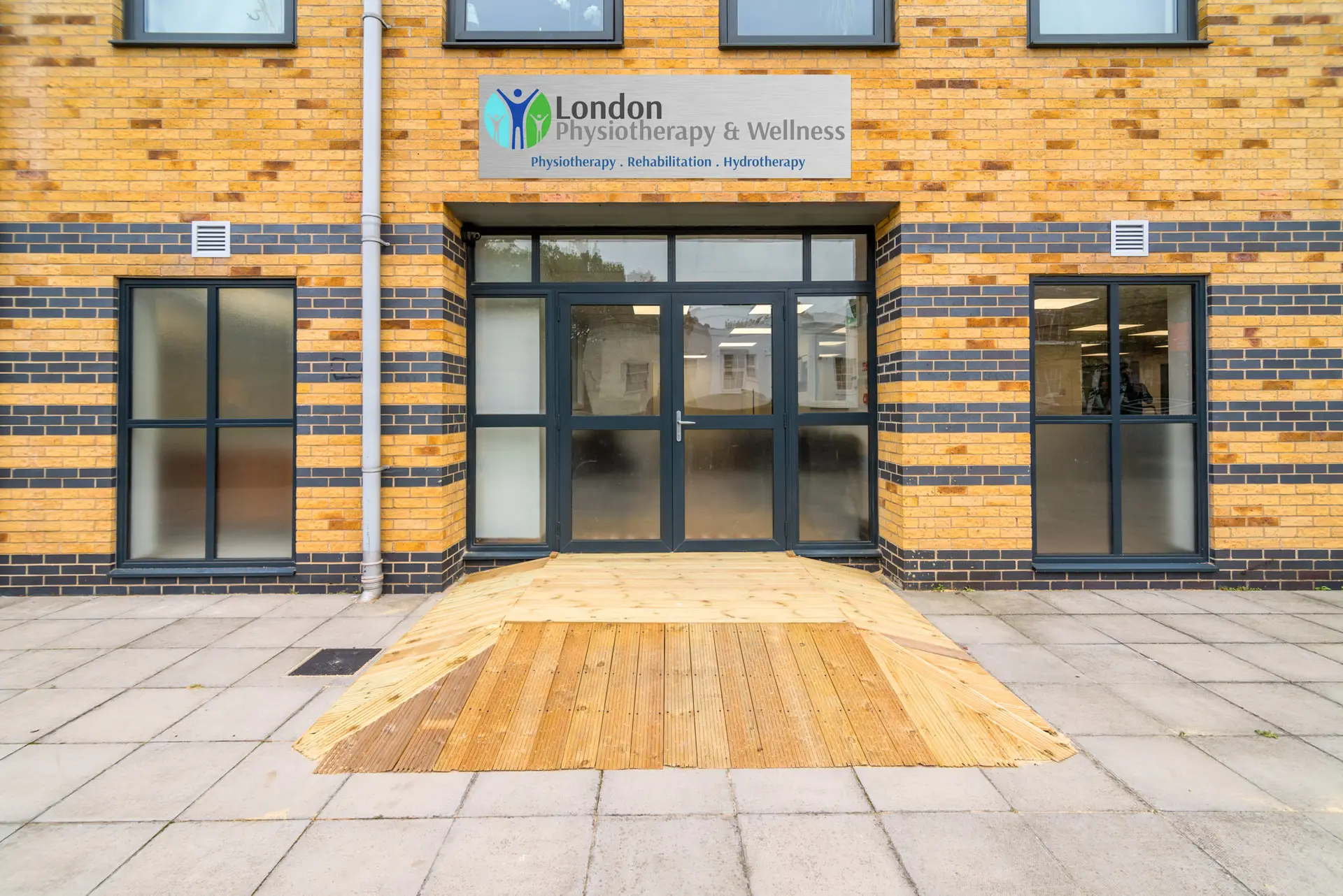
The main LPAW clinic is in Bow, E3, London, right next to The Bow Quarter. This bright and spacious clinic offers 4 treatment rooms, 2 changing rooms with showers, a large rehab gym, & onsite hydrotherapy in our 17 foot pool.
The LPAW satellite clinic is based in Stratford East Village where we run a thriving sports rehab offering.
Meniscus Tear Treatment in East London — Conservative Rehab & Post-Surgical Recovery
The menisci are two C-shaped wedges of fibrocartilage — the medial and lateral menisci — that sit between the femur and tibia within the knee joint. They perform several important functions:
Load distribution: The menisci increase the contact area between the joint surfaces, helping distribute force across a wider area and reducing peak stress on the articular cartilage by up to 50%
Joint stability: The medial meniscus, in particular, acts as a secondary stabiliser against forward movement of the tibia, working alongside the ACL
Shock absorption: The fibrocartilage structure helps absorb impact forces during walking, running, and jumping
Lubrication and nutrition: The menisci help distribute synovial fluid across the joint surfaces, supporting cartilage health
Because of these important functions, preserving meniscal tissue is now considered a priority whenever possible. Over the past decade, orthopaedic management has shifted away from routine early surgery and towards meniscal preservation where appropriate.
Acute traumatic tears: These typically occur during a twisting injury when the foot is planted and the knee is loaded. Symptoms usually begin suddenly and are often followed by swelling within 24–48 hours. These injuries are common in sports such as football, rugby, basketball, and skiing. The medial meniscus is injured more frequently than the lateral meniscus.
Degenerative (atraumatic) tears: In adults over the age of 35–40, meniscal tears can occur with relatively minor stress or even without a clear injury, as part of age-related degenerative change. Degenerative meniscal changes are extremely common. A 2014 study published in the New England Journal of Medicine found evidence of meniscal degeneration on MRI in 61% of adults over 50 with knee pain, but also in 32% of asymptomatic adults over 50. This is important because the presence of a meniscal tear on MRI does not necessarily mean it is the source of the patient’s pain.
The menisci are two C-shaped wedges of fibrocartilage — the medial and lateral menisci — that sit between the femur and tibia within the knee joint. They perform several important functions:
Load distribution: The menisci increase the contact area between the joint surfaces, helping distribute force across a wider area and reducing peak stress on the articular cartilage by up to 50%
Joint stability: The medial meniscus, in particular, acts as a secondary stabiliser against forward movement of the tibia, working alongside the ACL
Shock absorption: The fibrocartilage structure helps absorb impact forces during walking, running, and jumping
Lubrication and nutrition: The menisci help distribute synovial fluid across the joint surfaces, supporting cartilage health
Because of these important functions, preserving meniscal tissue is now considered a priority whenever possible. Over the past decade, orthopaedic management has shifted away from routine early surgery and towards meniscal preservation where appropriate.
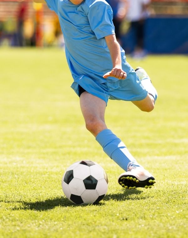
Acute traumatic tears: These typically occur during a twisting injury when the foot is planted and the knee is loaded. Symptoms usually begin suddenly and are often followed by swelling within 24–48 hours. These injuries are common in sports such as football, rugby, basketball, and skiing. The medial meniscus is injured more frequently than the lateral meniscus.
Degenerative (atraumatic) tears: In adults over the age of 35–40, meniscal tears can occur with relatively minor stress or even without a clear injury, as part of age-related degenerative change. Degenerative meniscal changes are extremely common. A 2014 study published in the New England Journal of Medicine found evidence of meniscal degeneration on MRI in 61% of adults over 50 with knee pain, but also in 32% of asymptomatic adults over 50. This is important because the presence of a meniscal tear on MRI does not necessarily mean it is the source of the patient’s pain.
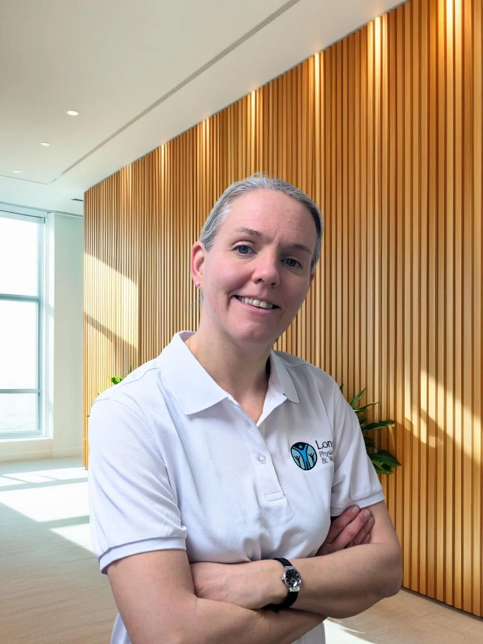
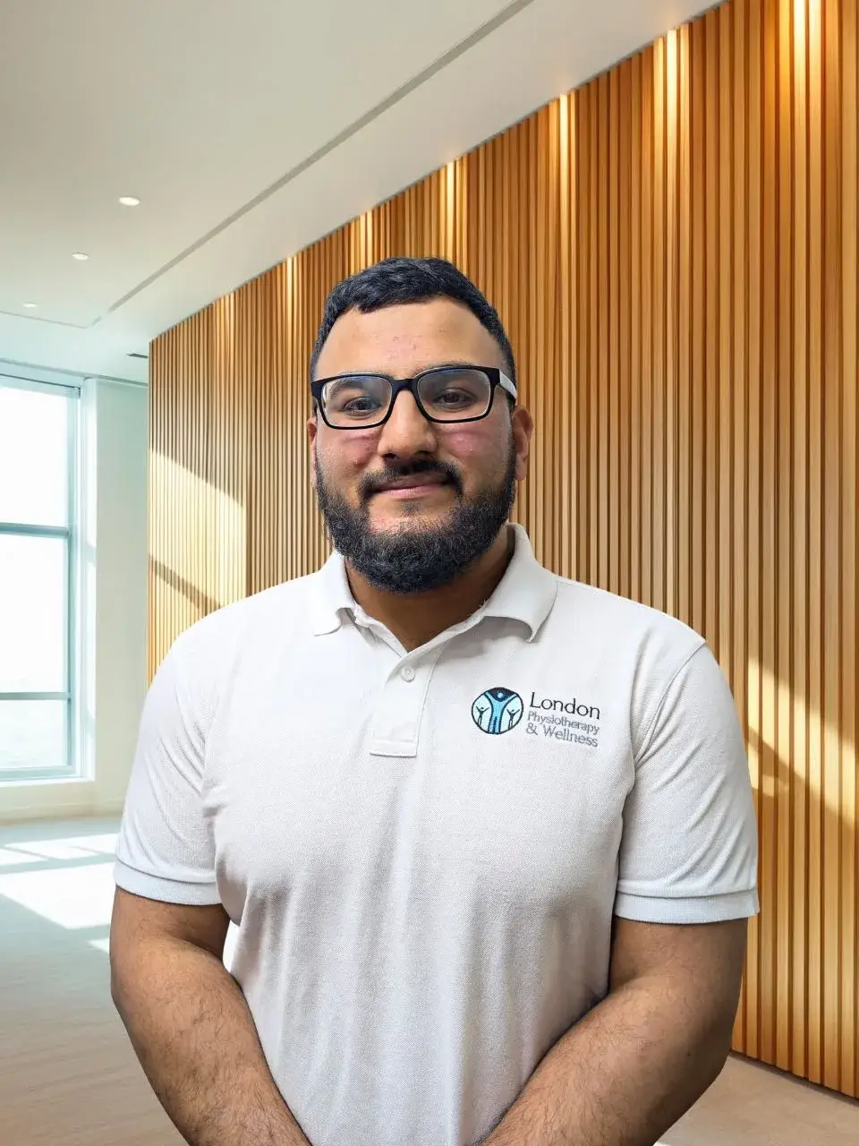
LPAW’s clinical team includes 19 practitioners, many holding postgraduate qualifications from UCL, King’s College London, and Guy’s and St Thomas’. Lead clinician Mr Arjun Viswanath MSc, MCSP, MPPA – Co-Founder and Consultant Physiotherapist – brings 25+ years of NHS and private experience including BMI London Independent Hospital and Harley Street.
Every clinician joining LPAW completes a mandatory intensive shadowing placement with our Consultant Physiotherapist before seeing patients independently. This is not a standard practice at most clinics – it’s our way of maintaining clinical consistency across the team.
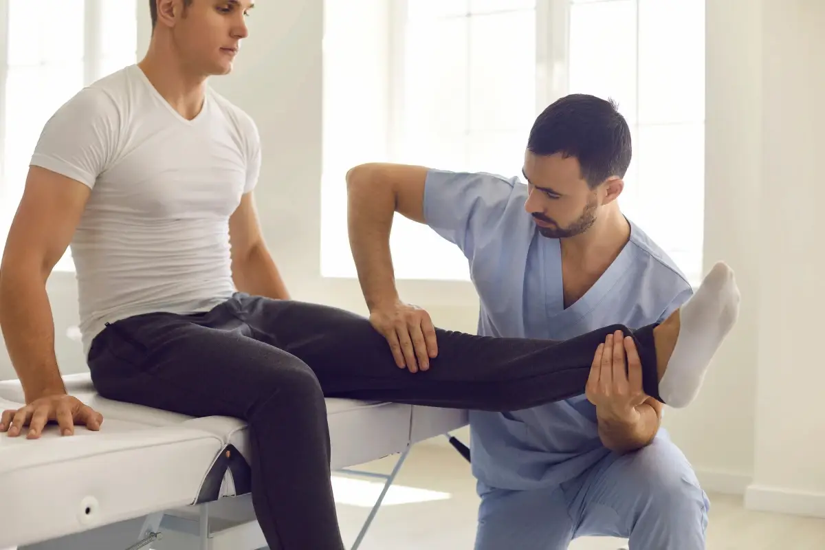
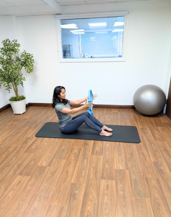
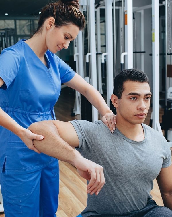
[Physiotherapy] for meniscus tears includes:
Quadriceps and hip strengthening: The foundation of conservative meniscus rehabilitation. Improving quadriceps strength can significantly reduce load through the meniscus and surrounding joint cartilage.
Range of motion restoration: Aimed at preventing joint stiffness while respecting pain levels and the specific tear pattern.
Load management: Gradual reintroduction of activities using a structured progression while monitoring symptom response.
Neuromuscular training: Exercises targeting balance, proprioception, and dynamic knee stability.
Activity modification guidance: Advice on temporarily avoiding aggravating activities such as deep squatting, twisting, and high-impact loading, while maintaining overall fitness during rehabilitation.
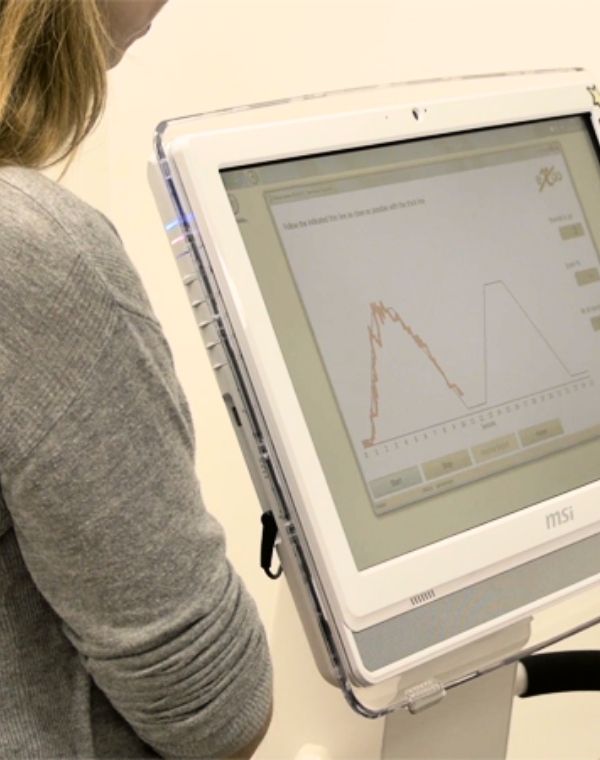
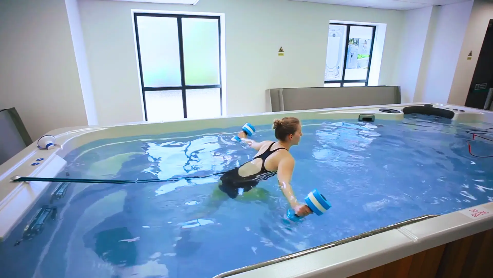
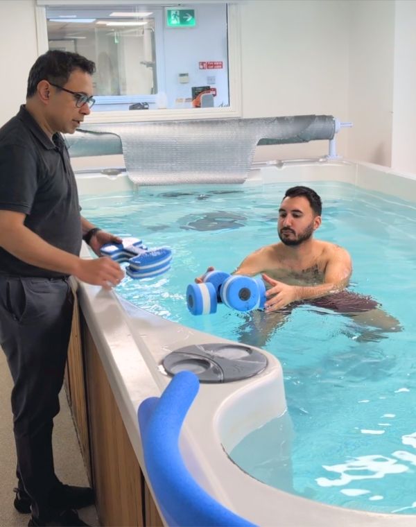
LPAW’s hydrotherapy pool — heated to 36°C — is invaluable for patients where back pain is severe enough to limit land-based exercise. The combination of buoyancy (reducing load on the spine) and warmth (reducing muscle spasm and improving circulation) allows movement and neural mobilisation exercises that are impossible or too painful on land. Hydrotherapy is particularly effective as an adjunct to Med-X rehabilitation for chronic back pain.
These are “red flags” that must be ruled out before physiotherapy treatment. Our physiotherapists conduct a full red flag screen at every initial assessment.
Following partial meniscectomy (removal of torn tissue): Return to full activity is generally faster than after meniscal repair, with most patients returning within 6–12 weeks. Rehabilitation focuses on restoring quadriceps function, regaining full range of motion, and progressively increasing load tolerance.
Following meniscal repair (suturing of the tear): Rehabilitation is more protective and gradual. Patients are commonly restricted to non-weight-bearing or toe-touch weight-bearing for approximately 4–6 weeks, often with limitations on knee flexion. Full rehabilitation typically takes 4–6 months. Hydrotherapy can be particularly useful during this phase because it allows earlier movement and loading in a reduced-impact environment.
In the long term, preserving meniscal tissue through repair is associated with better joint health outcomes compared with removing the damaged tissue.
LPAW’s hydrotherapy pool — heated to 36°C — is invaluable for patients where back pain is severe enough to limit land-based exercise. The combination of buoyancy (reducing load on the spine) and warmth (reducing muscle spasm and improving circulation) allows movement and neural mobilisation exercises that are impossible or too painful on land. Hydrotherapy is particularly effective as an adjunct to Med-X rehabilitation for chronic back pain.
Following partial meniscectomy (removal of torn tissue): Return to full activity is generally faster than after meniscal repair, with most patients returning within 6–12 weeks. Rehabilitation focuses on restoring quadriceps function, regaining full range of motion, and progressively increasing load tolerance.
Following meniscal repair (suturing of the tear): Rehabilitation is more protective and gradual. Patients are commonly restricted to non-weight-bearing or toe-touch weight-bearing for approximately 4–6 weeks, often with limitations on knee flexion. Full rehabilitation typically takes 4–6 months. Hydrotherapy can be particularly useful during this phase because it allows earlier movement and loading in a reduced-impact environment.
In the long term, preserving meniscal tissue through repair is associated with better joint health outcomes compared with removing the damaged tissue.


What We Do